All eukaryotic organisms contain cells that have a nucleus, organelles, and many internal membranes.
With all the wonderful diversity of life on Earth, however, you’re probably not surprised to discover that eukaryotic cells have many differences. By comparing the structure of a typical animal cell with that of a typical plant cell, you can see some of the differences among eukaryotic cells.- Cell walls, additional reinforcing layers outside the plasma membrane, are present in the cells of plants, fungi, and some protists, but not in animal cells.
- Chloroplasts, which are needed for photosynthesis, are found in the cells of plants and algae, but not animals.
- Large, central vacuoles, which contain fluid and are separated from the cytoplasm with a membrane, are found in the cells of plants and algae, but not animals.
- Centrioles, small protein structures that appear during cell division, are found in the cells of animals, but not plants.
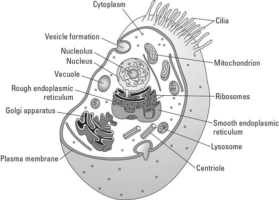 Structures in a typical animal cell
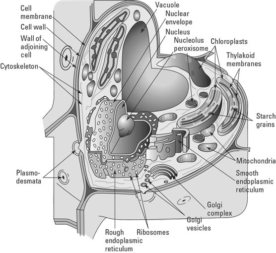 Structures in a typical plant cell
Home office: The nucleus
The nucleus houses and protects the cell’s DNA, which contains all of the instructions necessary for the cell to function. The DNA is like a set of blueprints for the cell, so you can think of the nucleus as the office where the blueprints are kept. If information from the blueprints is required, the information is copied into RNA molecules and moved out of the nucleus. The DNA plans stay safely locked away.The boundary of the nucleus is the nuclear envelope, which is made of two phospholipid bilayers similar to those that make up the plasma membrane.
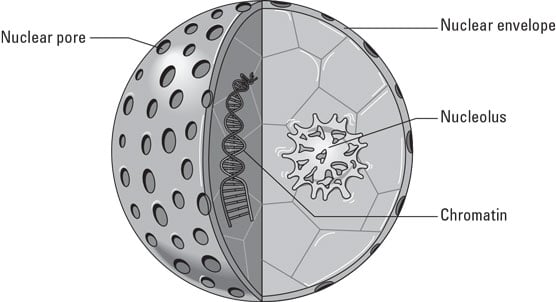 The nucleus
The phospholipids bilayers of the nuclear envelope are supported by a scaffold of protein cables, called the nuclear lamina, on the inner surface of the nucleus. The nuclear envelope separates the contents of the nucleus from the cytoplasm. The structures within the nucleus are
- DNA in the form of chromosomes or chromatin: When a cell is about to divide to make a copy of itself, it copies its DNA and bundles the DNA up tightly so that the cell can move the DNA around more easily. The tightly bundled DNA molecules are visible through a microscope as little structures in the nucleus called Most of the time, however, when a cell is just functioning and not about to divide, the DNA is very loose within the nucleus, like a bunch of long, very thin spaghetti noodles. When the DNA is in this form, it is called chromatin.
- Nucleoli where ribosomal subunits are made: Information in the DNA needs to be read in order to make the small and large subunits needed to build ribosomes. The cell builds the ribosomal subunits in areas of the nucleus called nucleoli. Then, the cell ships the subunits out of the nucleus to the cytoplasm, where they join together for protein synthesis. When you stain cells and look at them under the microscope, nucleoli look like large spots within the nucleus.
- RNA molecules and ribosomal subunits made in the nucleus must exit to the cytoplasm.
- Proteins made in the cytoplasm but needed for certain processes, such as copying the DNA, must cross into the nucleus.
- Nucleotides, building blocks for DNA and RNA, must cross into the nucleus so that the cell can make new DNA and RNA molecules.
- ATP molecules that provide energy for processes inside the nucleus like assembly of DNA molecules.
Traffic through the nuclear pores is controlled by proteins called importins and exportins. Proteins that are to be moved into or out of the nucleus have specific chemical tags on them that act like zip codes, telling the importins and exportins which way to move the protein with the tag. The movement of molecules into and out of the cell requires the input of energy from the cell in the form of adenosine triphosphate (ATP).
Post office: The endomembrane system
The endomembrane system, shown in the following figure, of the eukaryotic cell constructs proteins and lipids and then ships them where they need to go. Because this system is like a large package-shipping company, you can think of it as the post office of the cell.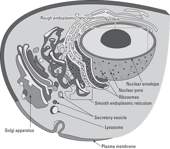 The endomembrane system
The endomembrane system has several components:
- The endoplasmic reticulum is a set of folded membranes that begins at the nucleus and extends into the cytoplasm. It begins with the outer membrane of the nuclear envelope and then twists back and forth like switchbacks on a steep mountain trail. The endoplasmic reticulum comes in two types:
- Rough endoplasmic reticulum (RER) is called rough because it’s studded with ribosomes. Ribosomes that begin to make a protein that has a special destination, such as a particular organelle or membrane, will attach themselves to the rough endoplasmic reticulum while they make the protein. As the protein is made, it’s pushed into the middle of the rough ER, which is called the Once inside the lumen, the protein is folded and tagged with carbohydrates. It will then get pushed into a little membrane bubble, called a transport vesicle, to travel to the Golgi apparatus for further processing.
- Smooth endoplasmic reticulum (SER) doesn’t have attached ribosomes. It makes lipids — for example, phospholipids for cell membranes. Lipids from the SER may also travel to the Golgi apparatus.
- The Golgi apparatus looks a little bit like a stack of pancakes because it’s made of a stack of flattened membrane sacs, called cisternae. The side of the stack closest to the nucleus is called the cis face of the Golgi, whereas the side farthest from the nucleus is called the trans Molecules arrive at the cis face of the Golgi and incorporate into the nearest cisterna. Lipids become part of the membrane itself, while proteins get pushed into the middle, or lumen, of the cisterna. The Golgi apparatus constantly changes as new cisternae form at the cis face, and old cisternae are removed from the trans face. As molecules make their journey through this flowing system, they’re modified and marked with chemical tags, so that they’ll get shipped to their proper destination.
- Vesicles are little bubbles of membrane in the cell and come in several types:
- Transport vesicles carry molecules around the cell. They’re like the large envelopes that you put your letters in. Transport vesicles travel from the ER to the Golgi and then to the plasma membrane to bring molecules where they need to go. They travel by gliding along protein cables that are part of the cytoskeleton.
- Lysosomes are the garbage disposals of the cell. They contain digestive enzymes that can break down large molecules, organelles, and even bacterial cells.
- Secretory vesicles bring materials to the plasma membrane so that the cell can release, or secrete, the materials.
- Peroxisomes are small organelles encircled by a single membrane. Often, they help break down lipids, such as fatty acids. Also, depending on the type of cell they are in, peroxisomes may be specialists in breaking down particular molecules. For example, peroxisomes in liver cells break down toxins, such as the ethanol from alcoholic beverages. In plants cells, glyoxisomes, a special kind of peroxisome, help convert stored oils into molecules that plants can easily use for energy.
Altogether, the endomembrane system works as a sophisticated manufacturing, processing, and shipping plant. This system is particularly important in specialized cells that make lots of a particular protein and then ship them out to other cells. These types of cells actually have more endoplasmic reticulum than other cells so that they can efficiently produce and export large amounts of protein.
As an example of how the endomembrane system functions, follow the pathway of synthesis and transport for an exported protein:- A ribosome begins to build a protein, such as insulin, that will be exported from the cell. At the beginning of the protein is a recognizable marker that causes the ribosome to dock at the surface of the rough endoplasmic reticulum.
- The ribosome continues to make the protein, and the protein is pushed into the lumen of the RER. Inside the lumen, the protein folds up, and carbohydrates are attached to it.
- The protein is pushed into the membrane of the RER, which pinches around and seals to form a vesicle, and the vesicle carries the protein from the RER to the Golgi.
- The vesicle fuses with the cis face of the Golgi apparatus, and the protein is delivered to the lumen of the Golgi, where the protein is modified.
- The protein eventually leaves in a vesicle formed at the trans face, which travels to the plasma membrane, fuses with the membrane, and releases the protein to the outside of the cell.
The fireplace: Mitochondria
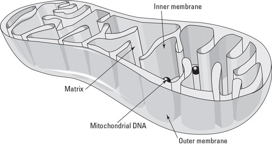
The mitochondrion (see the following figure) is the organelle where eukaryotes extract energy from their food by cellular respiration.Mitochondria are like the power plants of the cell because they transfer energy from food to ATP. ATP is an easy form of energy for cells to use, so mitochondria help cells get usable energy.
 The mitochondrion
Part of the process that extracts the energy from food requires a membrane, so mitochondria have lots of internal folded membrane to give them more area to run this process. Mitochondria actually have two membranes, the outer membrane and the inner membrane. The inner membrane is the one that is folded back and forth to create more area for energy extraction; the folds of this membrane are called cristae. The outer membrane separates the interior of the mitochondrion from the cytoplasm of the cell.
The two membranes of the mitochondrion create different compartments within the mitochondrion:
- The space between the two membranes of the mitochondrion is the intermembrane space.
- The inside of the mitochondrion is the
Mitochondria also contain ribosomes for protein synthesis and a small, circular piece of DNA that contains the code for some mitochondrial proteins. The ribosomes and DNA of mitochondria resemble those found in bacterial cells.
In the kitchen: Chloroplasts
Chloroplasts, shown in the following figure, are the place where eukaryotes make food molecules by the process of photosynthesis. Chloroplasts are found in the cells of plants and algae.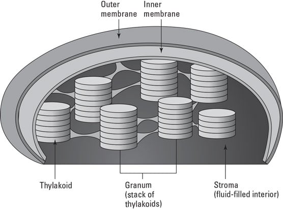 The chloroplast
Like mitochondria, chloroplasts have two membranes, an inner membrane and an outer membrane. In addition, they have little sacs of membranes called thylakoids stacked up in towers called grana.
The multiple membranes of the chloroplast divide it into several different spaces:
- The intermembrane space is between the inner and outer membranes.
- The central, fluid-filled part of the chloroplast is called the
- The interior of the thylakoid is another fluid-filled space.
Like mitochondria, chloroplasts contain their own ribosomes for protein synthesis and a small, circular piece of DNA that contains the code for some chloroplast proteins.
Scaffolding and railroad tracks: The cytoskeleton
The structure and function of cells are supported by a network of protein cables called the cytoskeleton, shown in the following figure. These proteins underlie membranes, giving them shape and support, much like scaffolding can support a building. Cytoskeletal proteins run like tracks through cells, enabling the movement of vesicles and organelles like trains on a railroad track. When cells swim by flicking whip-like extensions called cilia and eukaryotic flagella, they’re using cytoskeletal proteins. In fact, you use cytoskeletal proteins literally every time you move a muscle.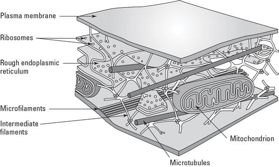 The cytoskeleton
Cytoskeletal proteins come in three main types, with each one playing a different role in cells:
- Microfilaments are made of the protein Microfilaments are the proteins that make muscle cells contract, help pinch animal cells in two during cell division, allow cells like amoebae to crawl, and act as railroad tracks for organelles in some types of cells.
- Microtubules are made of the protein tubulin. Microtubules are the proteins inside of cilia and flagella. They move chromosomes during cell division and act as railroad tracks for the movement of vesicles and some organelles.
- Intermediate filaments are made of various proteins. They often act as reinforcing proteins. For example, the protein lamin that strengthens the nuclear membrane is an intermediate filament. Likewise, the keratin that strengthens your skin cells and makes them resistant to damage is an intermediate filament.
You can easily mix up the words “microtubules” and “microfilaments.” Remember that “microtubules” are made of “tubul-in,” and they’re found in the “tube-shaped” cilia and flagella. (Okay, I’m stretching it on that last bit, but if it helps to remember it. . . .)
Motor proteins
Actin microfilaments and microtubules are long, cable-like proteins. They partner with motor proteins, proteins that use ATP to “walk” along the cables by repeatedly binding, changing shape, and releasing. Thus, the motor proteins use chemical energy to do cellular work in the form of movement. Several motor proteins work with microfilaments and microtubules:- Myosin often acts as a partner to actin. For example, when myosin walks along actin microfilaments in muscle cells, it causes the actin microfilament to slide. The sliding of actin microfilaments is what causes muscle contraction. Myosin also attaches to cellular components, such as chloroplasts in plant cells, and then walks along microfilaments. The movement of the motor proteins causes the cellular components to flow around the cell in a process called cytoplasmic streaming.
- Dynein partners with microtubules inside of cilia and eukaryotic flagella. When dynein walks along microtubules on one side of a cilium or flagellum, it causes the microtubules to bend. The bending of different parts of cilia and flagella makes them flick back and forth like little whips.
- Kinesin is another partner with microtubules. One end of the kinesin molecule attaches to vesicles, while the other end walks along the microtubules. The movement of kinesin causes the vesicles to slide along the microtubules like freight cars on a railroad track.
Cilia and flagella
Cilia and flagella are essentially the same structure, but cilia are typically shorter and more numerous on the surface of the cell whereas flagella are typically longer in length and fewer in number. Cilia are found on cells that make up the surfaces of tissues, such as cells in the respiratory and genital tracts of humans, where the cilia beat to move fluid and materials along the surface. For example, in the human respiratory tract, the beating of cilia moves mucus upward where you can cough it out of the body. Some cells, such as microscopic protists and sperm cells, swim using cilia and flagella.The internal structure of cilia and flagella is distinctive. If you cut a cilium or a flagellum crosswise and look at the circular end with an electron microscope, you’ll see the same pattern of microtubules in in both cilia and flagella, shown in the following figure. The microtubules are grouped in pairs, called doublets, that are similar to two drinking straws laid tightly together side by side.
The microtubules appear in a 9+2 arrangement, where nine pairs of microtubules (nine doublets) are arranged around the outside of the circle, while one pair of microtubules is in the center of the circle.
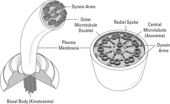 Structure of cilia and flagella
Rebar and concrete: Cell walls and extracellular matrices
The plasma membrane is the selective boundary for all cells that chooses what enters and exits the cell. However, most cells have additional layers outside of the plasma membrane. These extracellular layers provide additional strength to cells and may attach cells to neighboring cells in multicellular organisms. Typically, these layers are composed of long cables of carbohydrates or proteins embedded in a sticky matrix. The long, cable-like molecules work like rebar in concrete to create a strong substance. Two main types of extracellular layers support eukaryotic cells:- Cell walls are extra reinforcing layers that help protect the cell from bursting. Among eukaryotes, cell walls appear around the cells of plants, fungi, and many protists.
- The primary cell walls of plants and algae are made of cellulose. If the plant is a woody plant, lignin is also present. (Lignin is a complex molecule that hardens the cell walls of plants.)
- Fungal cell walls are made of chitin.
- The layer around animal cells is the extracellular matrix (ECM), shown in the following figure. This layer is made of long proteins, such as collagen, embedded in a polysaccharide gel. The ECM supports animal cells and helps bind them together. Animal cells actually attach themselves to the ECM via proteins, called integrins, that are embedded in the plasma membrane. The integrins bind to the actin microfilaments inside the cell and to ECM proteins called fibronectins that are outside the cell.
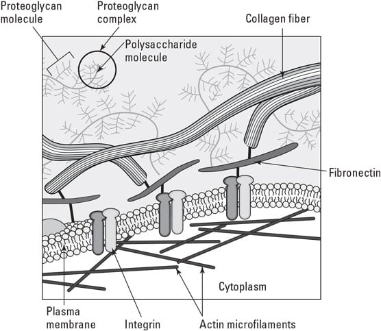 The extracellular matrix of animal cells