Neuroscience tells us that the neocortex interacts with the rest of the brain primarily through a structure called the thalamus. The thalamus, which is underneath (and hierarchically below) the neocortex, functions like a command center that controls what information goes between different parts of the neocortex and the rest of the brain.
While the neocortex can do very fine-grained analysis of the patterns you’re looking at, the thalamus controls where you look. When your neocortex is damaged, you lose particular skills. If your thalamus is damaged sufficiently, you lose consciousness.
The hypothalamus controls homeostatic body functions such as temperature and circadian rhythms.
Think of the thalamus as the gateway to the cortex. Virtually all signals from the senses are relayed through the thalamus, as are the signals from other subcortical areas. Many areas of the neocortex also communicate with each other through the thalamus.
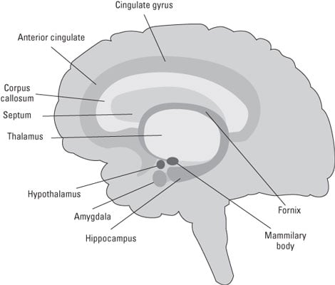
Below the neocortex and the thalamus are several important subcortical brain areas. One of the most important is a network of distinct, phylogenetically old nuclei called the limbic system. (Saying that these limbic system nuclei are phylogenetically old means that they existed in species much older than mammals, such as lizards, birds, and probably dinosaurs). Several important structures are within the limbic system:
The hippocampus: The hippocampus has a crucial function in the creation of memory. The hippocampus receives inputs from virtually the entire neocortex. Through specialized adjustable synaptic receptors called NMDA receptors, it can associate together virtually any constellation of properties that define an object and its context.
The amygdala: The amygdala is primarily involved with emotional processing and memory. The amygdala interacts with the prefrontal cortex to generate and process the major emotions of anger, happiness, disgust, surprise, sadness, and, particularly, fear. People who have sustained damage to their amygdalas have reduced abilities to react to and avoid situations that induce fear.
Orbitofrontal cortex: The orbitofrontal cortex is where the amygdala and other structures of the limbic system interact with the part of the prefrontal cortex. Suppose that, on some particular Friday evening while driving home, you’re almost hit by another car at a particular intersection. It is very likely that, for a long time after that, when approaching that intersection, particularly on Fridays, you’ll get a little twinge of fear or uneasiness. Your orbitofrontal cortex has stored the circumstances, and the amygdala has stored the fear.
The anterior cingulate cortex: The anterior cingulate cortex seems to monitor the progress toward whatever goal you’re pursuing and generates an “uh-oh” signal when things aren’t working out to indicate a change in strategy may be in order.
The basal ganglia: The basal ganglia consist of five major nuclei: the caudate, putamen, globus palladus, substantia nigra, and subthalamic nucleus. These nuclei comprise a highly interconnected system that interacts with the thalamus and neocortex to control behavior.