
With the explosion of genetic technology, there is a whole menu of options for detecting genetic abnormalities in your baby, and many of them can be done in the first trimester. The options fall mainly into two categories: screening tests and diagnostic tests.
A screening test will take bits of information about the developing fetus that can be collected and used to estimate the probability that your baby has certain genetic abnormalities and is usually expressed as a ratio (1/100, 1/1,000, 1/10,000, and so on). The bits of information can be substances produced by the placenta that can be measured in the mother’s blood, measurements on an ultrasound, or fragments of fetal DNA that circulate in mom’s bloodstream.
A diagnostic test utilizes tiny pieces of placental tissue (chorionic villus sampling, or CVS), fetal skin cells that have flaked off the surface of the baby (amniocentesis), or fetal lymphocytes (fetal blood sampling) to directly look at the tissue to see if the genetic makeup is normal or abnormal.
The screening tests available in the first trimester are the First Trimester Screen (utilizing components from the maternal blood and measurement of the nuchal translucency on the back of the baby’s neck by ultrasound).
The diagnostic tests involve testing the developing baby’s chromosomes. Chromosomes carry the genetic information (DNA) that determines what a person is like. People normally have 46 chromosomes — 23 inherited from their mother and 23 from their father. The 23rd pair of chromosomes are the sex chromosomes, which can be either XX (girl) or XY (boy).
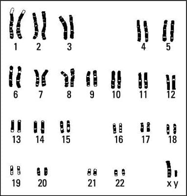
A woman has two X chromosomes, so she can only give an X chromosome to her offspring. A man has one X and one Y chromosome and can therefore give either to his offspring. As you can see, the man determines the baby’s sex, so you know who is to blame if you don’t get the little girl or little boy you were hoping for!
Certain abnormalities in chromosome number or structure can lead to problems in the baby. For example, Down syndrome, one of the more common chromosomal abnormalities associated with severe mental retardation, may occur if the fetus has an extra copy of chromosome 21. (The condition is also known as Trisomy 21, because the fetus has three copies of chromosome number 21.)
Looking at age
In the past, women who were going to be age 35 or older at their due date were offered the chance to undergo prenatal diagnosis to check the fetal chromosomes. Thirty-five was chosen as the target age because a woman’s risk of having a baby with a chromosomal abnormality increases significantly after she reaches that age.
It was also the age at which the risk of miscarriage from the procedure itself was equal to the actual chances that the fetus would have a chromosomal abnormality. However, although the risk of a chromosome abnormality is much less for women under the age of 35, most babies with Down syndrome are born to women under the age of 35 simply because they have more babies as a group than women older than 35.
The cutoff age of 35 is somewhat arbitrary and not followed in all countries. In Great Britain, for example, women are offered prenatal chromosomal testing at age 37 or after. Once nuchal translucency testing became available, the American College of Obstetricians and Gynecologists recommended that doctors stop using age to determine who should have a diagnostic test.
The current practice is to offer every woman the same thing and allow her to choose which test or tests she wants to undergo.
Even among women at risk for a chromosomal problem, some choose not to be tested, either because they don’t want to run any risk of miscarriage associated with the test or because of their personal beliefs about terminating a pregnancy.
Even if pregnancy termination isn’t something you would consider, prior knowledge of a fetus’s abnormalities can give you time to make preparations for a child that may have special needs.
Chorionic villus sampling
Chorionic villi are tiny, budlike pieces of tissue that make up the placenta. Because they develop from cells arising out of the fertilized egg, they have the same chromosomes and genetic makeup as the developing fetus.
By checking a sample of chorionic villi, the laboratory can see whether or not the chromosomes are normal in number and structure, determine the fetal sex, and test for some specific diseases (if the fetus may be at risk for these diseases).
Your doctor performs a chorionic villus sampling (CVS) by withdrawing placental tissue (containing chorionic villi) either through a hollow needle inserted through the abdomen (transabdominal CVS) or through a flexible catheter inserted through the cervix (transcervical CVS), depending on where the placenta is located within the uterus and the uterus’s general shape and position.
Your doctor uses ultrasound equipment as a guide as she performs the procedure. She then examines the tissue under a microscope, and the cells are cultured in a laboratory.
![[Credit: Kathryn Born, MA]](https://www.dummies.com/wp-content/uploads/438120.image1.jpg)
Like amniocentesis, CVS raises the risk of miscarriage slightly — about 1/1000. Neither CVS method is more risky than the other. The person performing the test should have plenty of experience doing the procedure; experience helps reduce the risk of miscarriage.
CVS results are typically available in seven to ten days. The main advantage that CVS has over amniocentesis is that it can provide information earlier in the pregnancy. This time factor may be important to some women who feel that termination is an option if severe abnormalities are present.
If you undergo CVS and are Rh-negative, you should receive an injection of Rh-immune globulin (Rhophylac or Rhogam are examples) following the procedure to prevent you from developing rH disease.