
No one test can absolutely detect multiple sclerosis (MS), but certain tests including magnetic resonance imaging (MRI) can be used to help confirm the diagnosis. The scan is a highly-sensitive, non-invasive way to view areas of damage in the central nervous system (CNS).
MRI uses a strong magnet, radio frequency pulses, and specialized computer software to create detailed pictures of your brain and spinal cord. A contrast material called gadolinium, delivered by intravenous infusion during the scan, is often used to highlight active areas of inflammation in the CNS caused by a breakdown in the blood-brain barrier (BBB). The BBB generally prevents bad stuff from passing from your bloodstream into your CNS.
As part of the diagnostic process, most neurologists want to see an MRI scan of the brain and spinal cord even though it can’t, by itself, determine if a patient has MS. Normal aging and other diseases besides MS can cause lesions (or, more informally, spots or scars) in the brain that look like MS. So, your doctor has to use other tools to ensure an accurate diagnosis.
Also, neurologists know that about 5 percent of people with definite MS can have a normal-appearing brain MRI scan, at least for a while, which means that scanning may need to be repeated in a few months in order to help confirm the diagnosis or rule it out. However, the longer the brain scan remains normal, the more doubtful the MS diagnosis becomes.
Different types of MRI scans yield different kinds of information. When studying your MRI images, the neurologist is looking for evidence of new disease activity as well as signs of prior attacks.
The telltale signs of prior attacks may include old lesions or damage to the axons, which are the parts of nerve cells that carry messages from one cell to another. MRI signs of damaged axons are called hypointensities or “black holes”. The doctor is also looking to find the total amount of damage that has occurred, which is referred to as lesion load.
Be sure to ask your neurologist to review your MRI images with you so that he or she can explain what kinds of changes have occurred in your brain or spinal cord and what these changes indicate about your MS. Because docs tend to use a lot of technical jargon when they talk about MRI scans, be sure to ask questions if you don’t understand the information you’re being given.
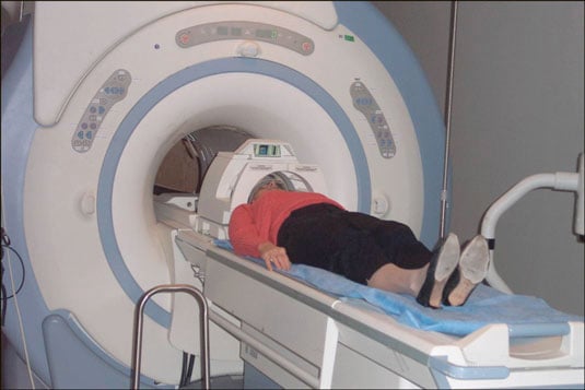
The figure shows a conventional MRI scanner. A conventional MRI scanner is an enclosed cylindrical magnet. During the scanning procedure you must lie perfectly still for several seconds to several minutes at a time, for about 45 minutes. If you have difficulty in enclosed spaces (for example, if you suffer from claustrophobia), the referring physician can give you a sedating medication prior to the scan. As an alternative, some MRI facilities offer machines with openings on the sides. To date, however, scans taken in open-sided machines don’t have the same degree of clarity as conventional scanners.
Because an MRI scanner uses strong magnets to obtain images of the body, patients who have cardiac pacemakers or certain kinds of metal implants can’t have this test. When scheduling the scan, the MRI office staff member will ask whether you have any prosthetic joints; a pacemaker; any implanted ports or infusion catheters; an intrauterine device (IUD); or any metal plates, pins, or screws from prior surgeries.
Not all metal causes a problem, so don’t assume that you can’t have an MRI just because you’ve had a pin or screw implanted sometime in the past. The staff member will also ask if you have any body tattoos or tattooed eyeliner (some people with tattoos report swelling or burning in the tattooed areas, and some tattoos contain metallic components that can interfere with image quality).
At the time of the scan, the MRI technician asks you to remove anything that may interfere with the quality of the images of your head, such as hairpins, jewelry, eyeglasses, hearing aids, and removable dental work. Because of the loud banging noise made by the scanner, the technician can offer you ear plugs or earphones to wear during the procedure.
If the gadolinium enhancement is required, the doctor inserts an intravenous needle into your vein. He or she then positions you comfortably (sort of) on a sliding table that rolls into the cylindrical tube. You can communicate with the technician at all times in the event that you need assistance.
Other than the discomfort you feel from having to lie perfectly still in a confined space with loud noises banging in your ears (sounds fun, huh?), the MRI procedure is painless.