The figure shows a diagram of a typical mechanoreceptor neuron.
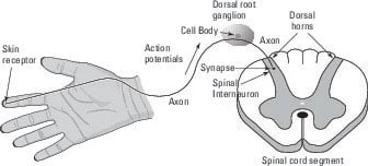
The cell bodies of somatosensory receptor neurons for most of our skin (below the head and neck) are located in a series of what are called ganglia (concentrations of neural cell bodies) just outside the dorsal root of the spinal cord. They are thus called dorsal root ganglia. These ganglia and the neurons they contain are part of the peripheral nervous system.
The cell bodies of the somatosensory neurons have no dendrites. Instead, a single axon leaves the cell body and then bifurcates, or separates into two paths, a short distance away.
One end of the axon enters the spinal cord at the dorsal root and makes conventional synapses on spinal interneurons, enabling the stretch reflex and the relaying somatosensory information to other spinal cord segments and up the spinal cord to the brain.
Here's the interesting part: The other end of the axon goes away from the dorsal root ganglion in a bundle with other axons and ends up in the skin, where it forms one of the receptor types: Merkel disk, Meissner corpuscle, Ruffini corpuscle, or Pacinian corpuscle.
The mechanoreceptors are activated directly when a mechanical force stimulates an axonal ending of one of these neurons. This activation occurs through a special ion channel that responds to the stretching of the membrane (in a typical neurotransmitter receptor, the activation is triggered by voltage or ligand binding). Here's what happens:
- The stretching causes action potentials to originate in the axonal ending and then proceed toward the cell body in the dorsal root ganglion (most axons conduct action potentials away from the cell body).
- The action potential continues past the axonal bifurcation point near the cell body into the spinal cord, where it reaches axon terminals and makes conventional synapses onto spinal interneurons.
- The interneurons connect the receptor neuron to motor neurons for reflexes and also send messages about the receptor activation to other spinal cord segments and up to the brain.